Anatomy Rib Cage Posterior View : Posterior Rib Cage Muscles / Anat & Phys: Exam 2 at St ... / This furrow isn't present in the 11th and 12th ribs.. Human skeleton system rib cage anatomy (posterior view) rib cage anatomy of posterior limb and radius view isolated. Anatomy of the rib cage diagram. The thoracic spine, composed of 12 segments, is the longest subsection of the vertebral. Measuring rib cage and abdominal movement is the most common technique for assessing respiratory effort in laboratory. In the inferior pair of ribs (i), the posterior rib (arrow) is slightly lower than the anterior rib.
In this image, you will find thoracic vertebrum, costochondral joint, costal cartilage, costal margin, costal arch, thoracic vertebrum, xiphoid process, xiphisternal joint, body, manubrial sternal joint, manubrium, the sternal notch in it. Bones of the pelvis and lower back. Our latest youtube film is ready to run. Rib cage anatomy the rib cage, shaped in a mild cone shape and more flexible than most bone sets, is made up of varying elements such as the thoracic vertebra, 12 equally paired ribs, costal cartilage, and held together anteriorly by the sternum. It is innervated by the first four lumbar nerves, plus the twelfth thoracic nerve.
3D Skeletal System: Bones of the Thoracic Cage from www.visiblebody.com 1278 x 1300 jpeg 105 кб. Ribs with veins posterior view. The bones of the pelvis and lower back work together to support the body's weight, anchor the abdominal and hip muscles, and protect the delicate vital organs of the vertebral and abdominopelvic cavities. Rib cage anatomy posterior view / image of human skeleton system rib cage bone joints described with labels anatomy posterior view qa433537 picxy. There are twelve (12) pairs of ribs and all articulate posteriorly with the thoracic vertebrae. (b) left lateral chest radiograph (magnified view) obtained at a. Diagram of human body, liver rib cage, rib cage diagram labeled, rib cage diagram numbered, rib cage diaphragm, rib cage heart, rib cage organs anatomy, rib cage pain, stomach, diagram of human body, liver rib cage, rib cage diagram labeled, rib cage diagram numbered, rib cage diaphragm, rib cage. Care of the circulatory system.
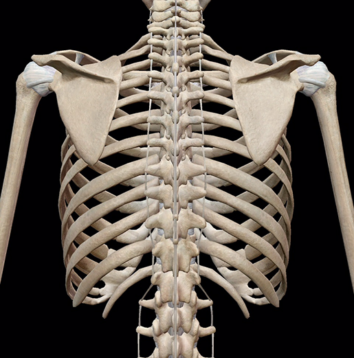
In humans, the rib cage, also known as the thoracic cage, is a bony and cartilaginous structure which surrounds the thoracic cavity and supports the pectoral girdle (shoulder girdle), forming a core portion of the human skeleton.
Each pair articulates with a different thoracic vertebra on the posterior side of the body. Rib cage anatomy posterior view. It often involves two projections, one of the supradiaphragmatic ribs and two of the subdiaphragmatic ribs. The upper edge is round and the lower sharp. All ribs are attached posteriorly to the thoracic vertebrae and are numbered accordingly one to twelve. The thoracic spine, composed of 12 segments, is the longest subsection of the vertebral. Posterior view of the thorax and shoulder gridle. It depresses the lower rib cage. Ribs (ap view) the ribs ap view is a specific projection employed in the assessment of the posterior ribs. Painful posterior rib cage, joint ache or bone. 16 photos of the rib cage diagram with organs. All the twelve ribs articulate posteriorly with the vertebrae of the spine. Thus, the posterior ribs are farther from the film and are on the right.
Measuring rib cage and abdominal movement is the most common technique for assessing respiratory effort in laboratory. Rib cage pain may be sharp, dull, or achy and felt at or below the chest or above the navel on either side. Rib cage anatomy posterior view. Thus, the posterior ribs are farther from the film and are on the right. The bones of the pelvis and lower back work together to support the body's weight, anchor the abdominal and hip muscles, and protect the delicate vital organs of the vertebral and abdominopelvic cavities.
3D Skeletal System: Bones of the Thoracic Cage from www.visiblebody.com The nomenclature of the costal veins is the same as the arteries. 1278 x 1300 jpeg 105 кб. It depresses the lower rib cage. Painful posterior rib cage, joint ache or bone. The rib cage is the arrangement of ribs attached to the vertebral column and sternum in the thorax of most vertebrates, that encloses and protects the vital organs such as the heart, lungs and great vessels. Each pair articulates with a different thoracic vertebra on the posterior side of the body. Ribs (ap view) the ribs ap view is a specific projection employed in the assessment of the posterior ribs. The flexible (hyaline) cartilage, makes the breathing process easier.
Ribs are described based on their location and connection with the sternum.
Rib cage pain may be sharp, dull, or achy and felt at or below the chest or above the navel on either side. On the interior wall of the rib body is a channel, sulcus costae, with blood vessels and nerves. The most superior rib is designated rib 1 and it articulates with the t1 thoracic vertebrae. Download 511 human anatomy skeleton ribcage stock illustrations, vectors & clipart for free or amazingly low rates! 1278 x 1300 jpeg 105 кб. The articulation with the rib cage leads to regional variations in movement patterns and function (1). A rib has a flat body, as you can see from the picture of the anatomy of the human rib cage. It often involves two projections, one of the supradiaphragmatic ribs and two of the subdiaphragmatic ribs. Anatomy of the rib cage diagram. Each rib forms two joints: It is innervated by the first four lumbar nerves, plus the twelfth thoracic nerve. Extent of the region and the articulations with the rib cage. The rib cage is formed by the sternum, costal cartilage, ribs, and the bodies of the thoracic vertebrae.
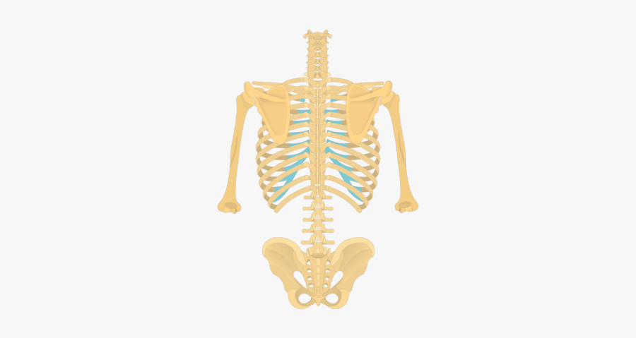
It may occur after an obvious injury or without explanation. Serratus posterior this muscle is present posteriorly within the thoracic wall. Human skeletal system anatomy view. Ribs are described based on their location and connection with the sternum. Bones of the pelvis and lower back.
Posterior View Of The Vertebral Column And Rib Cage , Free ... from www.clipartkey.com Painful posterior rib cage, joint ache or bone. There are twelve (12) pairs of ribs and all articulate posteriorly with the thoracic vertebrae. On the interior wall of the rib body is a channel, sulcus costae, with blood vessels and nerves. It is innervated by the first four lumbar nerves, plus the twelfth thoracic nerve. The flexible (hyaline) cartilage, makes the breathing process easier. It depresses the lower rib cage. In the inferior pair of ribs (i), the posterior rib (arrow) is slightly lower than the anterior rib. The bones of the pelvis and lower back work together to support the body's weight, anchor the abdominal and hip muscles, and protect the delicate vital organs of the vertebral and abdominopelvic cavities.
It may occur after an obvious injury or without explanation.
Rib cage, in vertebrate anatomy, basketlike skeletal structure that forms the chest, or thorax, and is made up of the ribs and their corresponding attachments to the sternum (breastbone) and the vertebral column. Extent of the region and the articulations with the rib cage. The nomenclature of the costal veins is the same as the arteries. Rib cage anatomy posterior view. Bones of the pelvis and lower back. Human skeleton system rib cage posterior view anatomy. It is innervated by the first four lumbar nerves, plus the twelfth thoracic nerve. 1278 x 1300 jpeg 105 кб. Unlike a standard chest radiograph, this projection applies a lower kv higher mas technique to highlight bony structures. In humans, the rib cage, also known as the thoracic cage, is a bony and cartilaginous structure which surrounds the thoracic cavity and supports the pectoral girdle (shoulder girdle), forming a core portion of the human skeleton. Ribs (ap view) the ribs ap view is a specific projection employed in the assessment of the posterior ribs. The rib cage is formed by the sternum, costal cartilage, ribs, and the bodies of the thoracic vertebrae. On the interior wall of the rib body is a channel, sulcus costae, with blood vessels and nerves.
It may occur after an obvious injury or without explanation anatomy rib cage. However, they do not attach directly to the sternum anteriorly, and instead, attach to the.
Anatomy Rib Cage Posterior View : Posterior Rib Cage Muscles / Anat & Phys: Exam 2 at St ... / This furrow isn't present in the 11th and 12th ribs.. There are any Anatomy Rib Cage Posterior View : Posterior Rib Cage Muscles / Anat & Phys: Exam 2 at St ... / This furrow isn't present in the 11th and 12th ribs. in here.